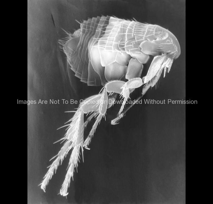
Electron Micrograph of a Parasitic Flea
Photos and electron micrographs of fleas
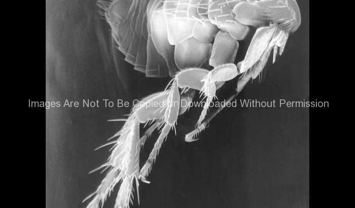
Electron Micrograph of a Parasitic Flea
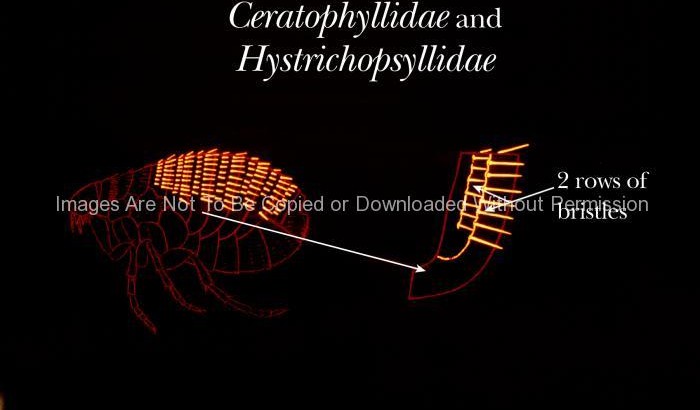
Note the difference in the arrangement of the tergal bristles on Ceratophyllidae and Hystrichopsyllidae fleas.
In general, fleas in the families Ceratophyllidae and Hystrichopsyllidae have two or more rows of bristles on each abdominal tergite, which are the plate-like, exoskeletal components of the flea’s abdomen.
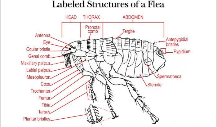
This illustration shows the identifying morphologic structures of an adult female flea.
Female fleas may be recognized by the presence of a spermatheca, which after mating, functions as a sperm storage receptacle. The characteristics of the spermatheca vary, i.e. size, shape and pigmentation, from specie to specie.
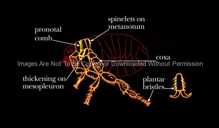
This illustration shows some common identifying characteristics found on the thorax of fleas.
Identifying features on the thorax include a pronotal comb, an internal thickening of the mesopleuron, spinelets on the metanotum, bristles/spinelets on the 1st segment (coxae) of the legs, and the plantar bristles on the last segment of the leg.
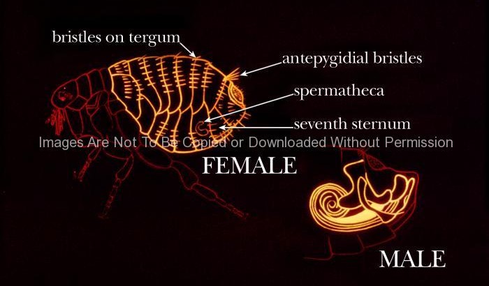
This illustration shows some common identifying characteristics found on the abdomen of the flea.
Identifying abdominal structures include the number of rows of tergal bristles, and number of antepygidial bristles; in females, the shape of the spermatheca and 7th sternal segment, in males, it’s the shape of certain structures in the terminal segment.
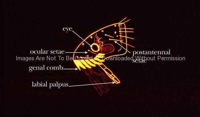
This diagram highlights some common identifying morphologic features found on the head region of fleas.
Identifying features include the absence or presence of an eye, the number and placement of ocular and post-antennal setae, the number of teeth in the genal comb, and the structure of the mouth parts, particularly the length of the labial palpi.
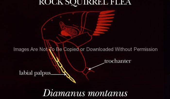
This illustration shows some of the identifying characteristics of the “rock squirrel” flea, Diamanus montanus.
D. montanus, has a very long pair of labial palpi, extending beyond the tip of the trochanter of its first pair of legs. This flea is known to be a plague vector in the United States, associated with rock squirrels, Spermophilus variegatus.
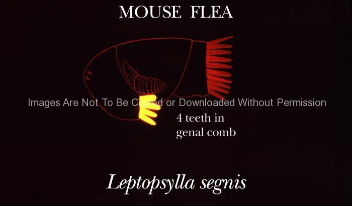
This illustration shows one of the identifying morphologic characteristics of the “mouse flea”, Leptopsylla segnia.
Leptopsylla segnia has 4 prominent teeth in the genal comb on either side of its head; it also lacks eyes. It is more closely associated with rats of the genus Rattus spp. than with mice. It has been found to be a poor plague vector.
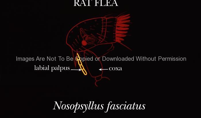
This illustration reveals an identifying characteristic of the “rat flea’s”, Nosopsyllus fasciatus, head region.
N. fasciatus has a labial palpus that doesn’t extend beyond the tip of the first coxae. It’s closely associated with rats of the genus Rattus spp., and important in the transfer of plague from rat to rat, but doesn’t readily bite humans.
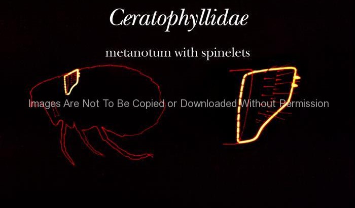
p>The posterior margin of the metanotum of fleas in the family Ceratophyllidae has spinelets, or tiny pigmented teeth.
Fleas that are members of the family Ceratophyllidae have been shown to be important in the transmission, and perpetuation of plague by acting as vectors in the transmission of Yersinia pestis bacteria amongst wild and domestic rodent hosts.