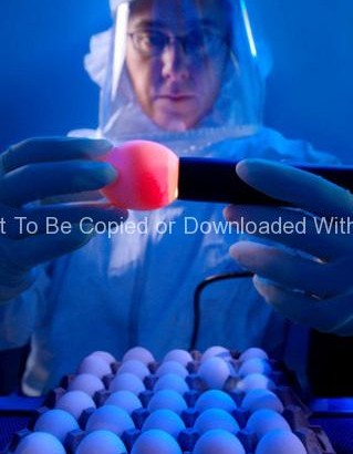
This 2008 photograph depicted CDC microbiologist, Amanda Balish, as she was demonstrating how one properly “candles” an embyonated chicken egg, which employs a very bright light that is either placed behind the egg, such as was done throughout history by using a candle, hence the name, or employing more modern methods, using a powerful lamp placed against the broad end of the egg, as was the case here. In this way, the contents of the egg are revealed through the translucent shell. By using this procedure, Amanda was able to access the viability of each egg used in the isolation of influenza viruses.
If the egg is unfertilized the observer will see only a round yolk sac, and no developing embryo, or any network of blood vessels. Unfertilized eggs are known as “yolkers”. Eggs known as “quitters”, had been fertilized, but the embryo had stopped growing. In the case of quitters, one will note a thin blood ring encircling the yolk, as seen in PHIL 10148. “Winners” are eggs that had been fertilized, and lead to the growth of a healthy embryo, which is seen in PHIL 10149. In this case, one will observe numbers of networked blood vessels surrounding the yolk, and depending upon the length of gestation, one might see a dark shadow representing the developing embryonic eye.