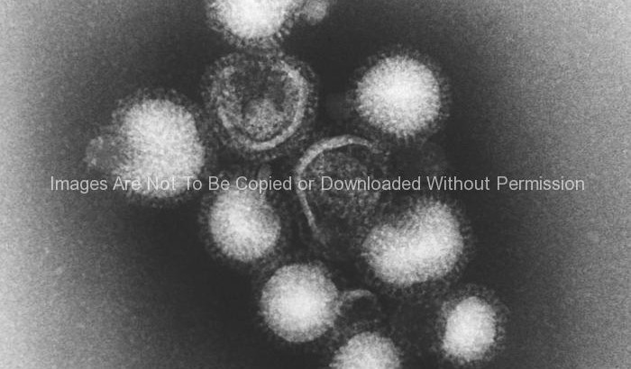
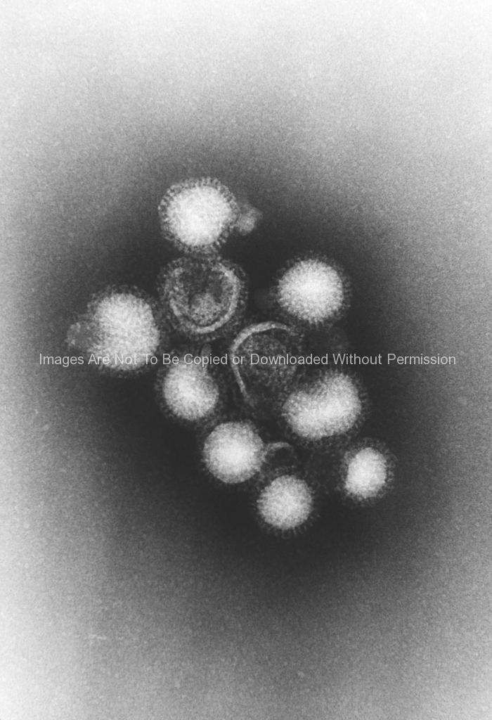
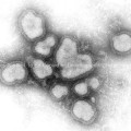
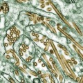
This negatively-stained transmission electron micrograph (TEM) depicted a small grouping of a number of influenza A virions, which also revealed at this very high magnification, the neuraminidase and hemagglutinin “spikes” protruding from the virions’ proteinaceous capsid coats. At its core, the genome consists of eight single-stranded segments of a negative-sense RNA ((-) ssRNA). Influenza A virions can be observed as being both spherical and filamentous in nature. Each strand is enclosed in, or “encapsidated” inside the viral nucleoprotein, thereby, forming what is known as the ribonucleoprotein (RNP) particle.
Other Photos You May Like:
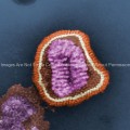
 Ultrastructural Details of an Influenza Virus Particle
 Ultrastructural Details of an Influenza Virus Particle
 Ultrastructural Details of an Influenza Virus Particle
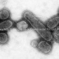 Micrograph of Recreated 1918 Influenza Virions
 Influenza A Virions
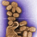 1918 Influenza Virions
 Influenza A Virions
 Electron Micrograph of Avian Influenza A