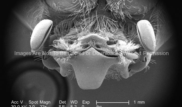
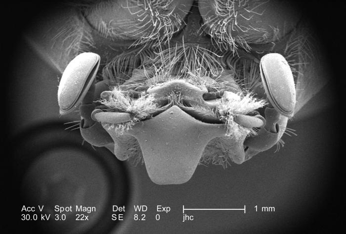
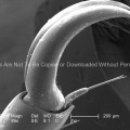
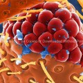
Equipped with “lamellate” type antennae, the distal three sensorial segments have been folded over one another, which can be seen here in this image. This morphology is typically found in members of the family Scarabaeidae.
The antennae provide the insect with data indicative of changes encountered in its environment such as chemical, thermal, and tactile queues. This beetle was found in the Decatur, Georgia suburbs.
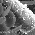
The dorsum of the adult figeater beetle is a drab green color, while its ventral surface is a vivid iridescent green. Their diet mainly consists of soft, overly ripe fruits including figs, hence its name. However, it’s the larval phase of this insect, which wrecks more havoc upon agricultural crops than adults, for the larvae, which burrow into the soils, will feed upon the crop roots, thereby, killing the fruit-bearing plants.
Also, note the numerous sensorial hair-like structures adorning the insect’s head, which are known as “setae”. These are not hairs in the mammalian sense, which are composed of keratin, but are exoskeletal adnexae, composed of chitin, as is the exoskeleton itself. These setae provide the figeater with information indicative of changes within its immediate environment such as thermal, tactile, and chemical fluctuations.
Other Photos You May Like:
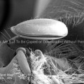 Distal Tip of This Adult “Figeater” Beetle’s, Cotinis mutabilis, Antenna.
 Exoskeletal Morphologic Characteristics (head of a beetle)
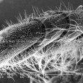
 Distal End of an Adult “figeater” Beetle’s, Cotinis mutabilis Leg
 Ultrastructural Morphology on the Head Region of a Larval Antlion
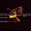 Head Region of Fleas Diagram
 Ultrastructural Morphology on the Rostral Head Region of a Bedbug (Cimex lectularius)
 Ultrastructural Morphology on the Head of a Bedbug (Cimex lectularius)
 Ultrastructural Morphology on the Head of a Bedbug (Cimex lectularius)