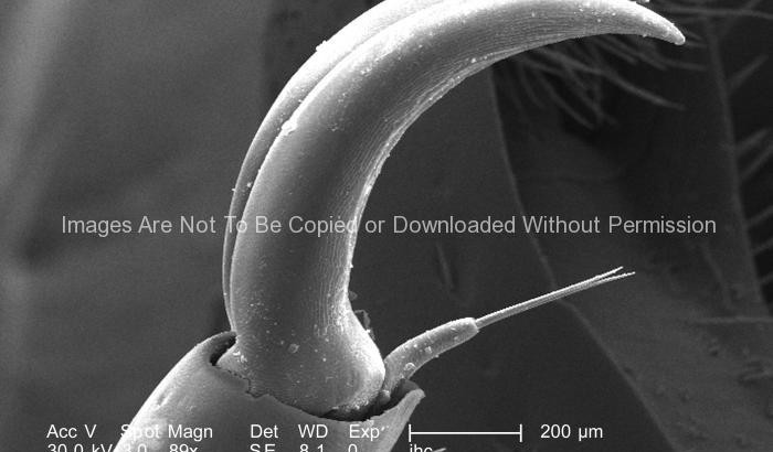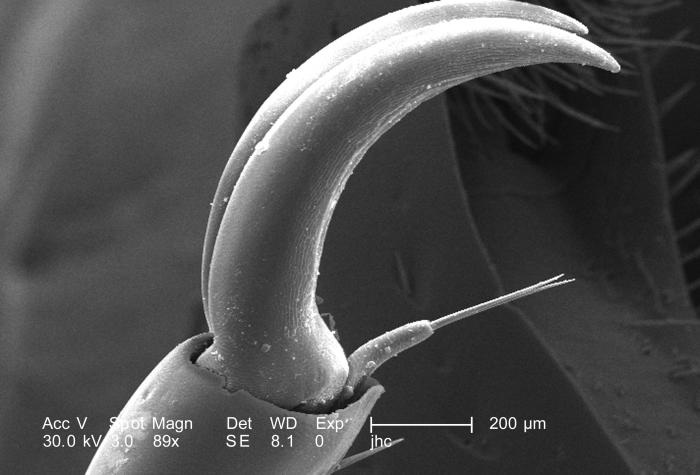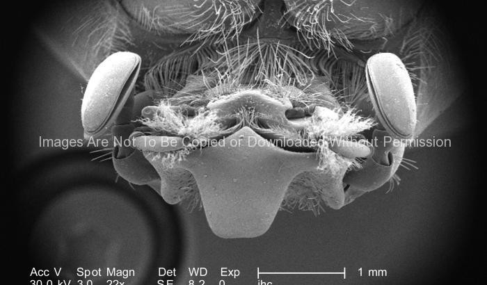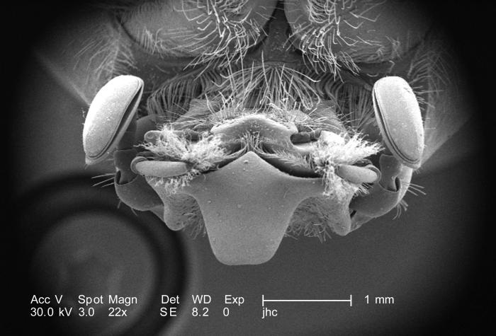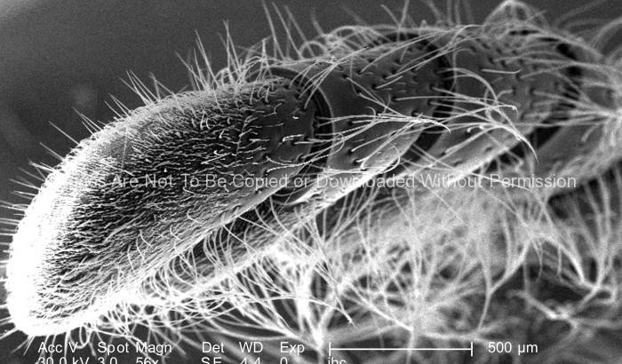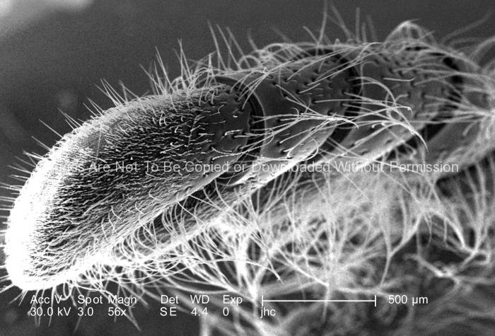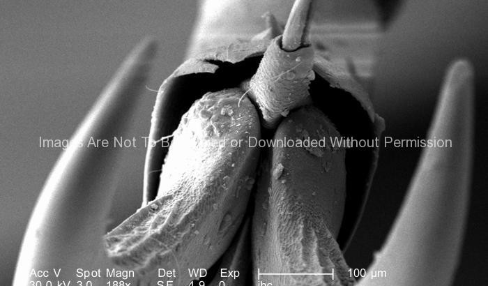
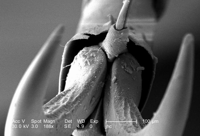
At a magnification of 188X, this scanning electron micrograph (SEM) depicted a head-on view of the distal clawed tip of an adult “figeater” beetle’s, Cotinis mutabilis leg. The insect leg is comprised of a variable number of segments, however, there are usually six which predominate, including the most proximal coxa, i.e., attaching the leg to the thorax, followed by the trochanter, femur, tibia, tarsus, and pretarsus, which in the case of this beetle is a claw with its spiked empodium.
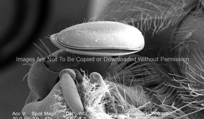
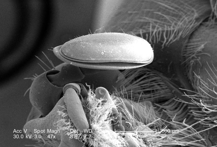
Due to the jointed nature of this organism’s leg configuration, it is classified in the phylum, Arthropoda, i.e., “Arthro” = jointed, and “poda” = legs. This beetle was found in the Decatur, Georgia suburbs.
The dorsum of the adult figeater beetle is a drab green color, while its ventral surface is a vivid iridescent green. Their diet mainly consists of soft, overly ripe fruits including figs, hence its name. However, it’s the larval phase of this insect, which wrecks more havoc upon agricultural crops than adults, for the larvae, which burrow into the soils, will feed upon the crop roots, thereby, killing the fruit-bearing plants.