11_819_lores1

Exoskeletal Surface of a Larval Staged Antlion

Exoskeletal Surface of a Larval Staged Antlion

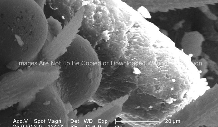
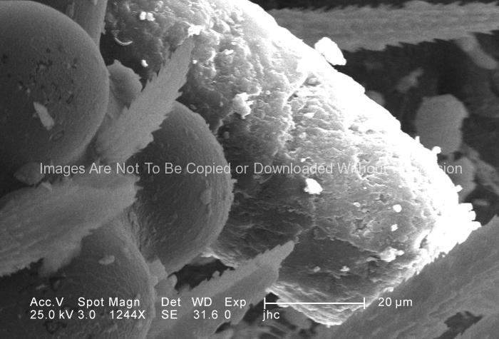
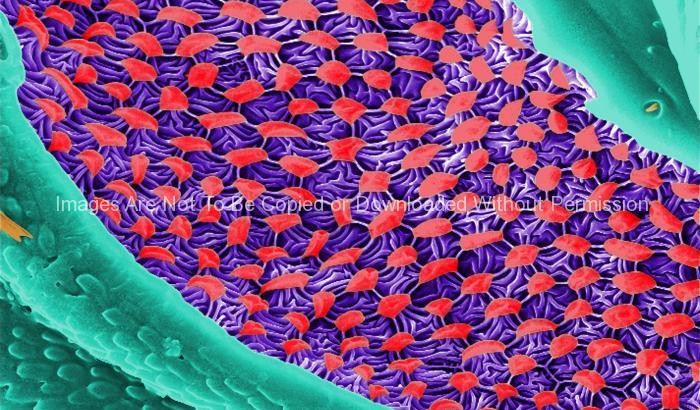
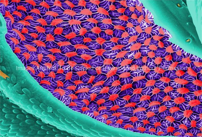
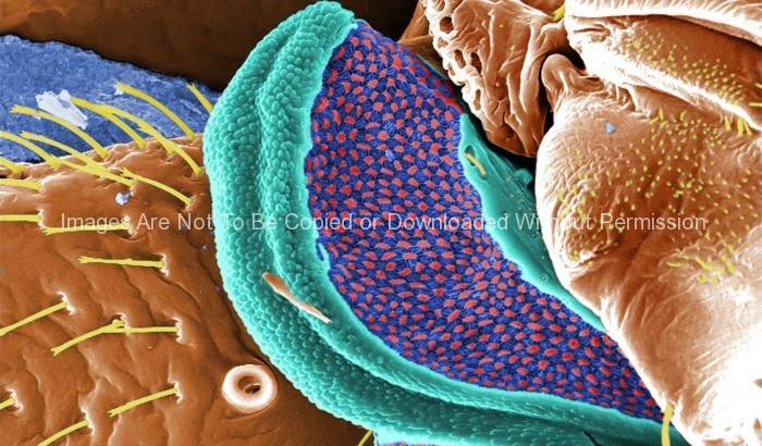
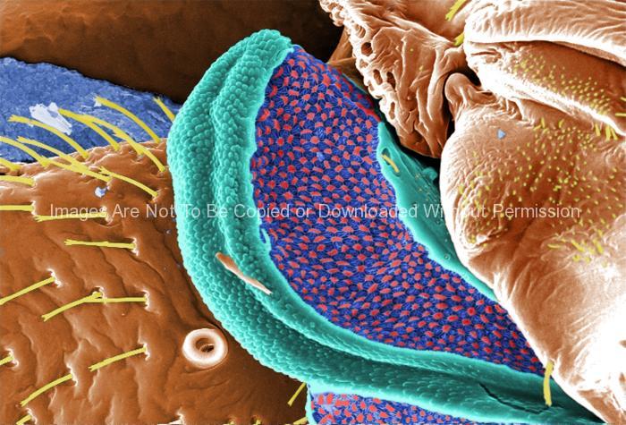
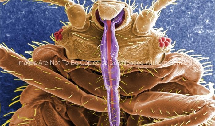
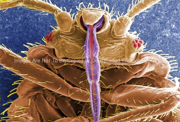
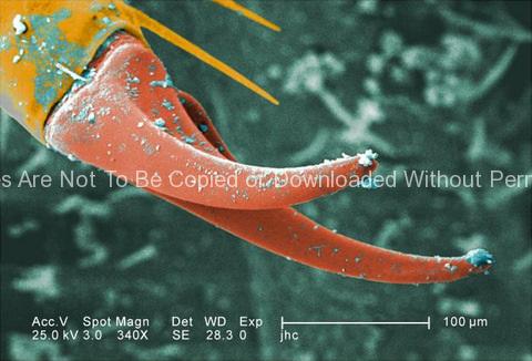
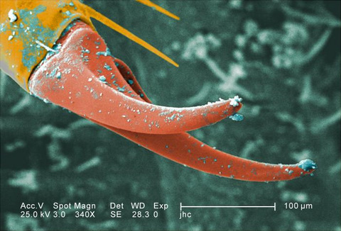
Under a moderate magnification of 340X, this digitally-colorized scanning electron micrograph (SEM) depicted the exoskeletal surface of a larval staged antlion, sometimes referred to as a “doodlebug”, because of the trails it leaves in the soft sand as it hunts for prey. This arthropod, i.e., jointed legs, undergoes dramatic morphologic changes when it metamorphoses into a beautiful flying antlion lacewing. In this particular view, the distal end of one of the larva’s extremities is highlighted, revealing the claw it uses to grasp objects it encounters in its environment, including sand.

Ultrastructural Morphology on the Head Region of a Larval Antlion

Ultrastructural Morphology on the Head Region of a Larval Antlion

Ultrastructural Morphology on the Head Region of a Larval Antlion

Under a magnification of 1244X, this scanning electron micrograph (SEM) depicted some of the ultrastructural morphology displayed on the head region of a larval antlion, surrounding the region of its right compound eye. This larva is sometimes referred to as a “doodlebug”, because of the trails it leaves in the soft sand as it hunts for prey. This arthropod, i.e., jointed legs, undergoes dramatic morphologic changes when it metamorphoses into a beautiful flying antlion lacewing. Note the particulate debris dispersed over the exoskeletal surface, which represents sand, and other constituents of the antlion’s natural environment.

11_793_lores1

Doodlebug (uncolorized)

Doodlebug (uncolorized)

Under a very low magnification of 13X, this scanning electron micrograph (SEM) depicted the entire ventral surface of a larval-staged antlion, sometime referred to as a “doodlebug”, because of the trails it leaves in the soft sand as it hunts for prey. This arthropod, i.e., jointed legs, undergoes dramatic morphologic changes when it metamorphoses into a beautiful flying antlion lacewing.

Doodlebug

Doodlebug

Under a very low magnification of 13X, this digitally-colorized scanning electron micrograph depicted the entire ventral surface of the larval staged antlion, sometime referred to as a “doodlebugs”, because of the trails they leave in the soft sand as they hunt for prey. These arthropods, i.e., jointed legs, undergo dramatic morphologic changes when it metamorphoses into a beautiful flying antlion lacewing.