1960s Nurse Handling Test Tubes

1960s Nurse Handling Test Tubes

1960s Nurse Handling Test Tubes

Ms. Beverly Lawrence, former Administrative Assistant in the Respiratory Virus Infections Unit at the Centers for Disease Control and Prevention, was shown here in this 1968 photograph handling test tubes containing frozen throat swab specimens that were to be tested for influenza.

Because the symptoms of influenza can be similar to those caused by other infectious agents, the disease can be difficult to diagnose based on clinical symptoms alone. A number of tests can help in the diagnosis of influenza, but testing does not need to be done for all patients. During an outbreak of respiratory illness, testing for influenza can help determine if influenza is the cause of the outbreak.

Researcher Using a Pipetter

Researcher Using a Pipetter

Researcher Using a Pipetter

This Centers for Disease Control microbiologist was shown here delivering tissue culture specimens into a microtiter plate using an instrument know as a pipetter. The culture samples were to be tested for the presence of the influenza virus.

The presence or absence of influenza virus would be determined by using an agglutination test. If it involved the agglutination, or clumping of red blood cells (RBCs), the phenomenon is known as hemoagglutination.

Diagnostic tests available for influenza include viral culture, serology, rapid antigen testing, polymerase chain reaction (PCR), and immunofluorescence assays. Sensitivity and specificity of any test for influenza might vary by the laboratory that performs the test, the type of test used, and the type of specimen tested. Among respiratory specimens for viral isolation or rapid detection, nasopharyngeal specimens are typically more effective than throat swab specimens. As with any diagnostic test, results should be evaluated in the context of other clinical and epidemiologic information available to health-care providers.

Two People on a Dog Sled in Alaska 7776_lores

Two People on a Dog Sled in Alaska

Two People on a Dog Sled in Alaska

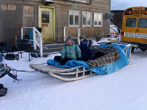
Laura Hammitt, M.D. (left), and Kim Boyd Hummel, R.N., B.S.N. (right) were photographed prior to getting on their way to the remote Native Alaska village of Teller, Alaska, in order to investigate an outbreak of invasive Haemophilus influenzae serotype b (Hib) meningitis.

This trip was made via dogsled, in order to conduct a review of regional immunization records, immunize children at risk for Hib, and to obtain nasal-pharyngeal swabs of village residents.

The trip was conducted under the auspices of the National Center for Infectious Diseases (NCID), Arctic Investigations Program. Drs. Hammitt and Butler were members of the CDC’s Epidemic Intelligence Service (EIS).

Teller, Alaska is north of Nome. The final destination was Brevig Mission (pop. 234), but due to dense ice fog, the plane went as far as Teller, dropped the team off, and turned around. They got a ride on this sled across the frozen inlet from Teller to Brevig Mission. The ride was 6 miles, which took about 20 minutes. The temperature varied between 20 and 25 degrees below zero.

Male Body Louse (Pediculus humanus var. corporis)

Male Body Louse (Pediculus humanus var. corporis)

Male Body Louse (Pediculus humanus var. corporis)

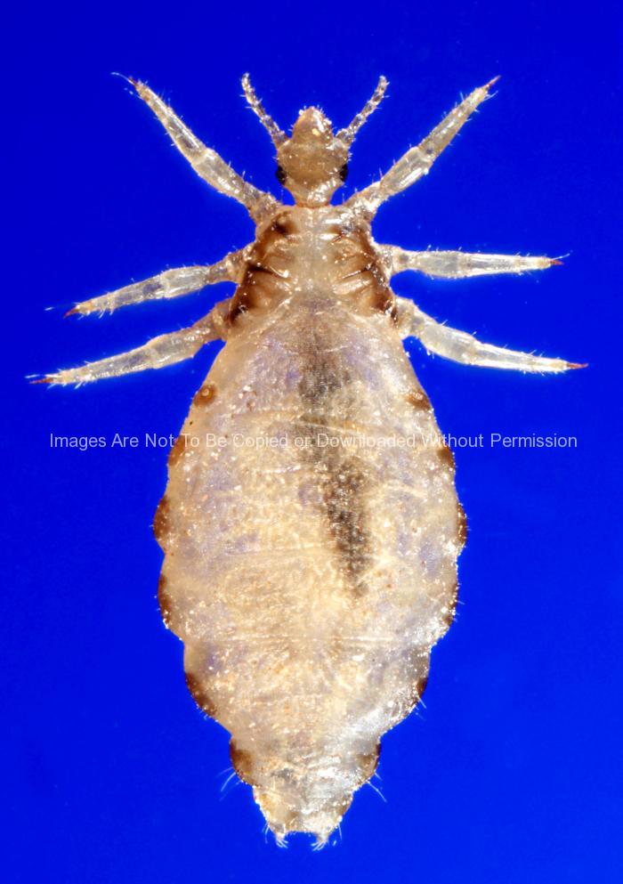
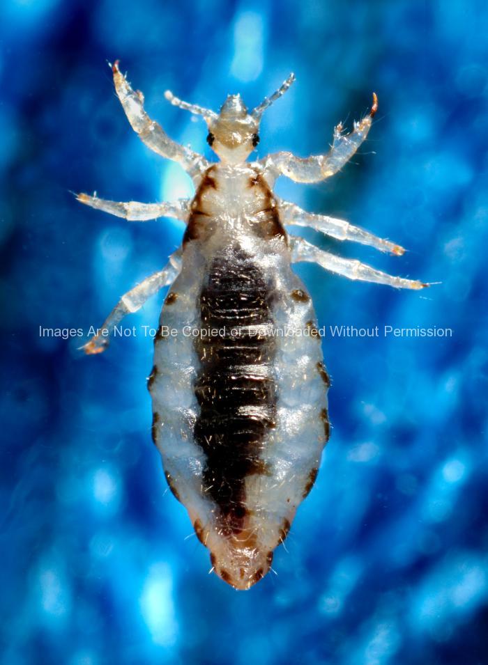
This photograph depicts a dorsal view of a male body louse, Pediculus humanus var. corporis. Some of the external morphologic features displayed by members of the genus Pediculus include an elongated abdominal region without any processes, and three pairs of legs, all equal in length and width. The distal tip of the male’s abdomen is rounded, whereas, the female’s is concave.

Body lice are parasitic insects that live on the body, and in the clothing or bedding of infested humans. Infestation is common, found worldwide, and affects people of all races. Body lice infestations spread rapidly under crowded conditions where hygiene is poor, and there is frequent contact among people. Note the sensorial setae, or hairs that cover the louse’s body, which pick up, and transmit information to the insect about changes in its environment such as temperature, and chemical queues. The dark mass inside the abdomen is a previously ingested blood meal.

Dorsal View of a Female Body Louse (Pediculus humanus var. corporis)

Dorsal View of a Female Body Louse (Pediculus humanus var. corporis)

Dorsal View of a Female Body Louse (Pediculus humanus var. corporis)

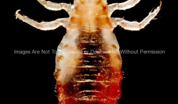
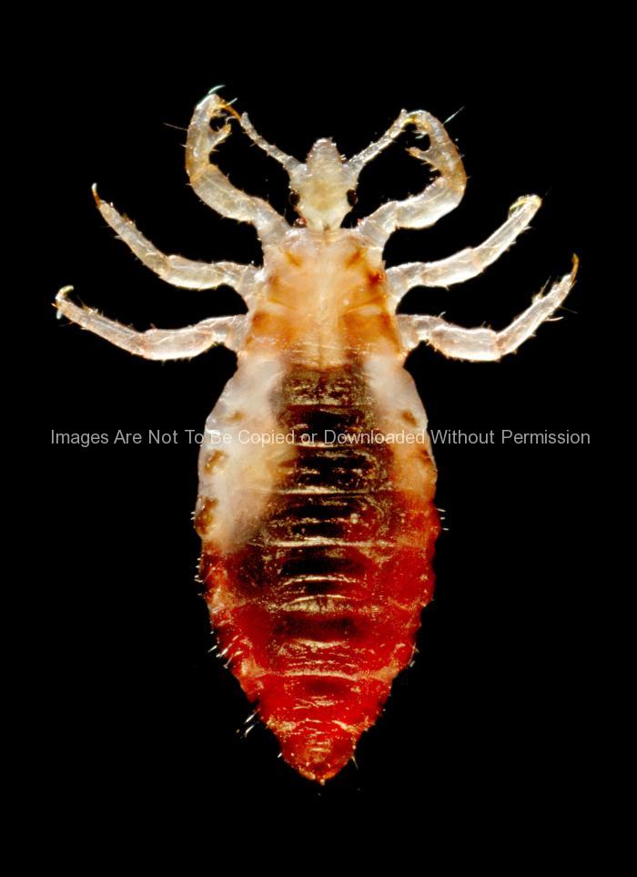
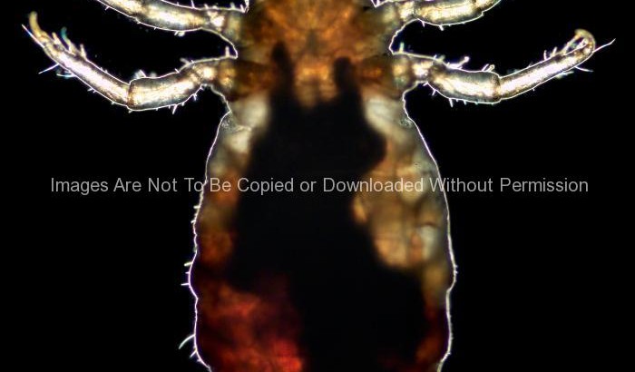
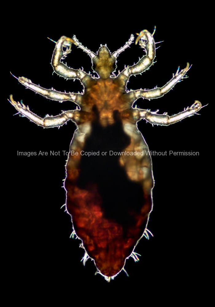
This photograph depicts a dorsal view of a female body louse, Pediculus humanus var. corporis. Some of the external morphologic features displayed by members of the genus Pediculus include an elongated abdominal region without any processes, and three pairs of legs, which are all equal in length and width. One of the anatomical features displayed by the female louse is its concave distal abdominal tip.

Body lice are parasitic insects that live on the body, and in the clothing or bedding of infested humans. Infestation is common, found worldwide, and affects people of all races. Body lice infestations spread rapidly under crowded conditions where hygiene is poor, and there is frequent contact among people.

How is a body lice infestation diagnosed?

By looking closely in the seams of clothing and on the body for eggs and for crawling lice. Diagnosis should be made by a health care provider if you are unsure about infestation.

Lateral View of a Female Body Louse (Pediculus humanus var. corporis)

Lateral View of a Female Body Louse (Pediculus humanus var. corporis)

Lateral View of a Female Body Louse (Pediculus humanus var. corporis)

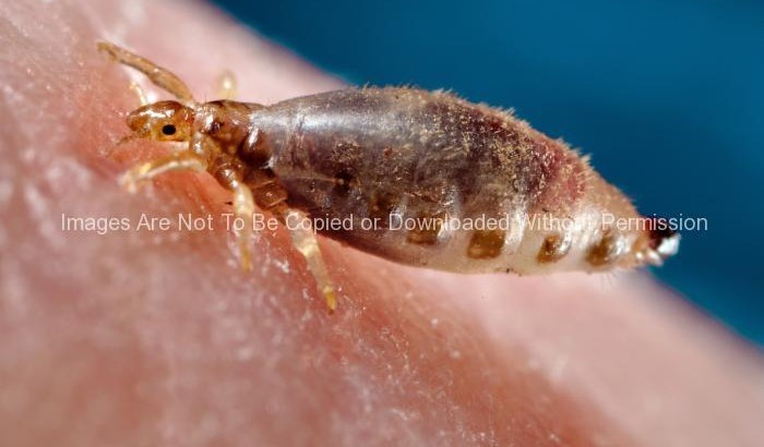
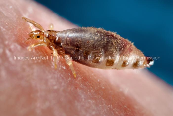
This photograph depicts a lateral view of a female body louse, Pediculus humanus var. corporis, as it was obtaining a blood-meal from a human host, who in this case, happened to be the photographer. Note its elongated abdominal region without any processes, and three pairs of legs, which are all equal in length and width, features displayed by Pediculus members.

Body lice are parasitic insects that live on the body, and in the clothing or bedding of infested humans. Infestation is common, found worldwide, and affects people of all races. Body lice infestations spread rapidly under crowded conditions where hygiene is poor, and there is frequent contact among people. Note the sensorial setae, or hairs that cover the louse’s body, which pick up, and transmit information to the insect about changes in its environment such as temperature, and chemical queues. The dark mass inside the abdomen is a previously ingested blood meal.

Are body lice infestations common in the United States?

Body lice are found only in homeless, transient populations who don’t have access to changes of clothes or bath. Infestation is unlikely to persist on anyone who bathes regularly and who regularly has access to freshly laundered clothing and bedding.

Dorsal View of a Male Body Louse (Pediculus Humanus Var. Corporis)

Dorsal View of a Male Body Louse (Pediculus Humanus Var. Corporis)

Dorsal View of a Male Body Louse (Pediculus Humanus Var. Corporis)

This photograph depicted a dorsal view of a male body louse, Pediculus humanus var. corporis. Some of the external morphologic features displayed by members of the genus Pediculus include an elongated abdominal region without any processes, and three pairs of legs, all equal in length and width. The distal tip of the male’s abdomen is rounded, whereas, the female’s is concave.

Body lice are parasitic insects that live on the body, and in the clothing or bedding of infested humans. Infestation is common, found worldwide, and affects people of all races. Body lice infestations spread rapidly under crowded conditions where hygiene is poor, and there is frequent contact among people. Note the sensorial setae, or hairs that cover the louse’s body, which pick up, and transmit information to the insect about changes in its environment such as temperature, and chemical queues. The dark mass inside the abdomen is a previously ingested blood meal.

What do body lice look like?

There are three forms of body lice: the egg (sometimes called a nit), the nymph, and the adult.

Nit: Nits are body lice eggs. They are generally easy to see in the seams of clothing, particularly around the waistline and under armpits. They are about the size of this mark ( ’ ). Nits may also be attached to body hair. They are oval and usually yellow to white. Nits may take 30 days to hatch.

Nymph: The egg hatches into a baby louse called a nymph. It looks like an adult body louse, but is smaller. Nymphs mature into adults about 7 days after hatching. To live, the nymph must feed on blood.

Adult: The adult body louse is about the size of a sesame seed, has 6 legs, and is tan to grayish-white. Females lay eggs. To live, adult lice need to feed on blood. If the louse falls off of a person, it dies within 10 days.

Ventral View of a Male Body Louse (Pediculus humanus var. corporis)

Ventral View of a Male Body Louse (Pediculus humanus var. corporis)

Ventral View of a Male Body Louse (Pediculus humanus var. corporis)

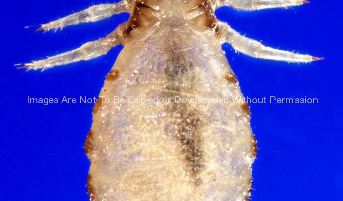
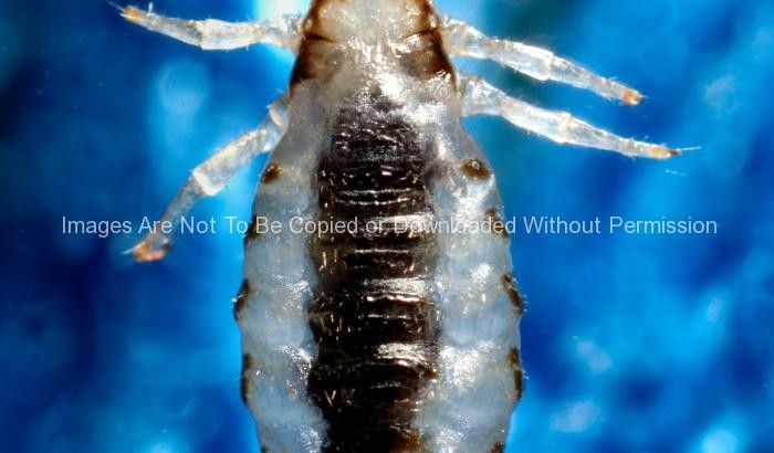
This photograph depicts a ventral view of a male body louse, Pediculus humanus var. corporis. Some of the external morphologic features displayed by members of the genus Pediculus include an elongated abdominal region without any processes, and three pairs of legs, all equal in length and width. The distal tip of the male’s abdomen is rounded, whereas, the female’s is concave.

Body lice are parasitic insects that live on the body, and in the clothing or bedding of infested humans. Infestation is common, found worldwide, and affects people of all races. The dark mass inside the abdomen is a previously ingested blood meal, obtained by the louse when the photographer offered his arm to the insect on which it fed.

How is a body lice infestation diagnosed?

By looking closely in the seams of clothing and on the body for eggs and for crawling lice. Diagnosis should be made by a health care provider if you are unsure about infestation.

9948_lores

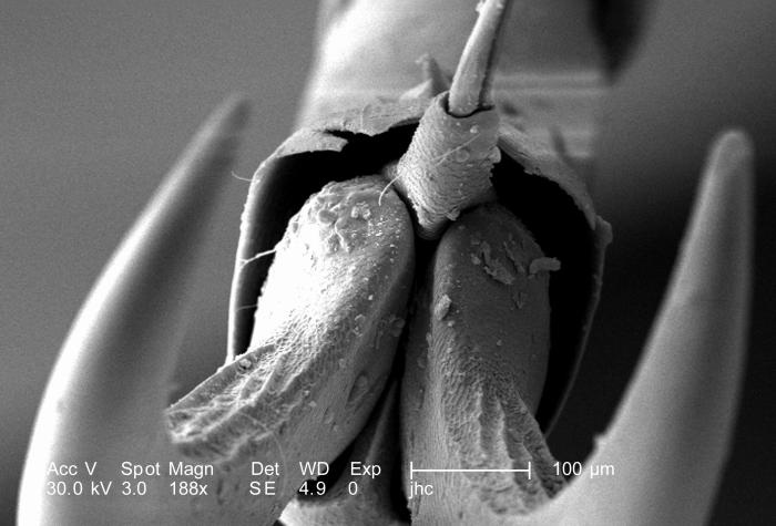
Distal Clawed Tip of an Adult “figeater” beetle’s, Cotinis mutabilis Leg

Distal Clawed Tip of an Adult “figeater” beetle’s, Cotinis mutabilis Leg

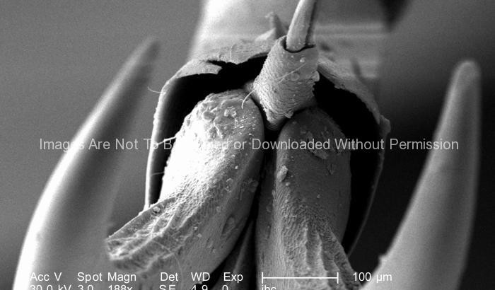
At a magnification of 188X, this scanning electron micrograph (SEM) depicted a head-on view of the distal clawed tip of an adult “figeater” beetle’s, Cotinis mutabilis leg. The insect leg is comprised of a variable number of segments, however, there are usually six which predominate, including the most proximal coxa, i.e., attaching the leg to the thorax, followed by the trochanter, femur, tibia, tarsus, and pretarsus, which in the case of this beetle is a claw with its spiked empodium.

Due to the jointed nature of this organism’s leg configuration, it is classified in the phylum, Arthropoda, i.e., “Arthro” = jointed, and “poda” = legs. This beetle was found in the Decatur, Georgia suburbs.

The dorsum of the adult figeater beetle is a drab green color, while its ventral surface is a vivid iridescent green. Their diet mainly consists of soft, overly ripe fruits including figs, hence its name. However, it’s the larval phase of this insect, which wrecks more havoc upon agricultural crops than adults, for the larvae, which burrow into the soils, will feed upon the crop roots, thereby, killing the fruit-bearing plants.