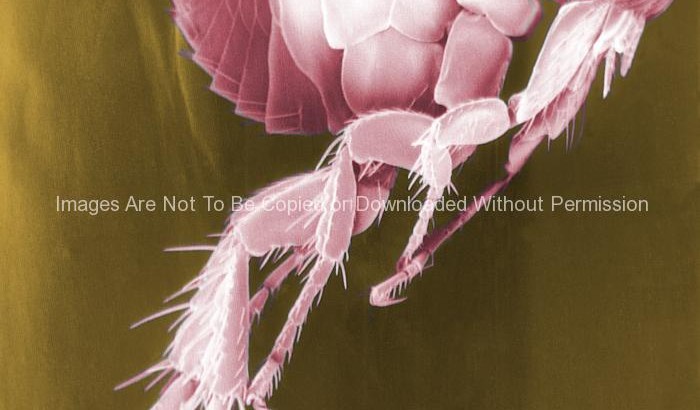
Scanning Electron Micrograph of a Flea. Fleas are known to carry a number of diseases that are transferable to human beings through their bites. Included in this infections is the plague, caused by the bacterium Yersinia pestis.
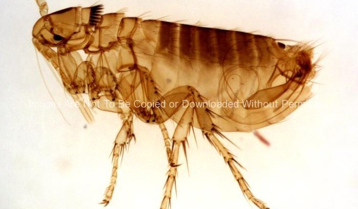
Adult male Oropsylla Montana flea, formerly known as Diamanus Montana.
This photograph depicts an adult male Oropsylla Montana flea, formerly known as Diamanus Montana.
Fleas are known to carry a number of diseases that are transferable to human beings through their bites. Included in this infections is the plague, caused by the bacterium Yersinia pestis.
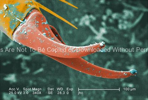
Exoskeletal Surface of a Larval Staged Antlion
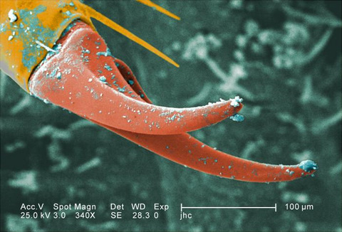
Under a moderate magnification of 340X, this digitally-colorized scanning electron micrograph (SEM) depicted the exoskeletal surface of a larval staged antlion, sometimes referred to as a “doodlebug”, because of the trails it leaves in the soft sand as it hunts for prey. This arthropod, i.e., jointed legs, undergoes dramatic morphologic changes when it metamorphoses into a beautiful flying antlion lacewing. In this particular view, the distal end of one of the larva’s extremities is highlighted, revealing the claw it uses to grasp objects it encounters in its environment, including sand.
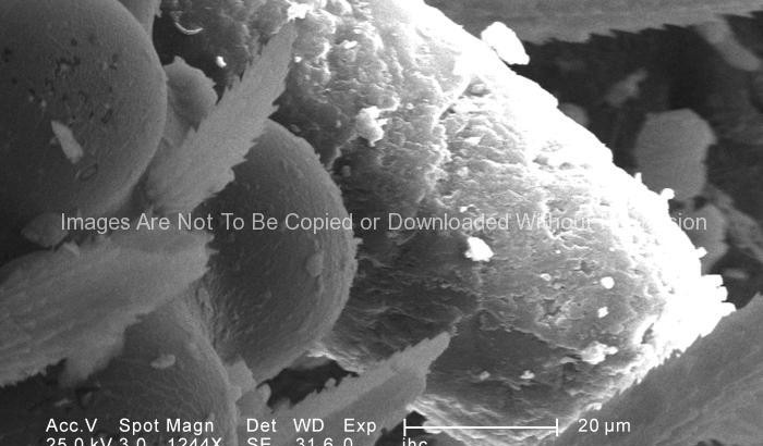
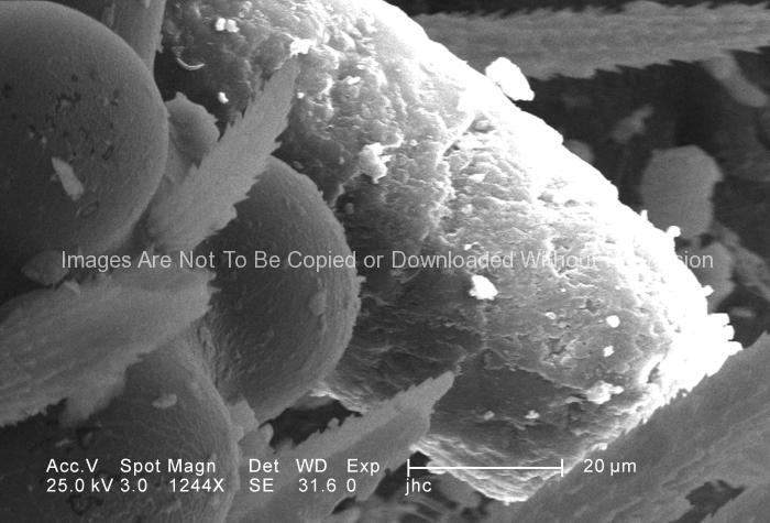
Ultrastructural Morphology on the Head Region of a Larval Antlion
Under a magnification of 1244X, this scanning electron micrograph (SEM) depicted some of the ultrastructural morphology displayed on the head region of a larval antlion, surrounding the region of its right compound eye. This larva is sometimes referred to as a “doodlebug”, because of the trails it leaves in the soft sand as it hunts for prey. This arthropod, i.e., jointed legs, undergoes dramatic morphologic changes when it metamorphoses into a beautiful flying antlion lacewing. Note the particulate debris dispersed over the exoskeletal surface, which represents sand, and other constituents of the antlion’s natural environment.
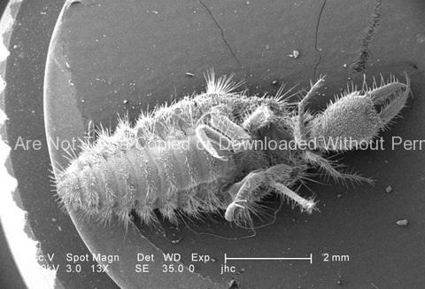
Doodlebug (uncolorized)
Under a very low magnification of 13X, this scanning electron micrograph (SEM) depicted the entire ventral surface of a larval-staged antlion, sometime referred to as a “doodlebug”, because of the trails it leaves in the soft sand as it hunts for prey. This arthropod, i.e., jointed legs, undergoes dramatic morphologic changes when it metamorphoses into a beautiful flying antlion lacewing.
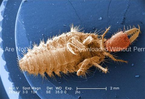
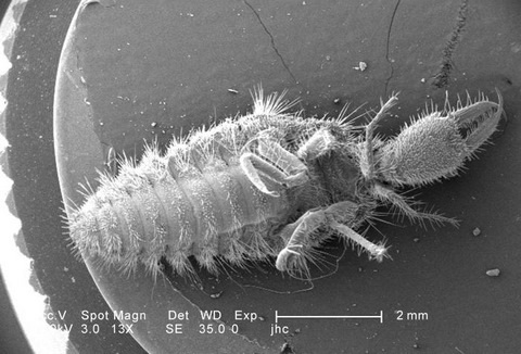
Doodlebug
Under a very low magnification of 13X, this digitally-colorized scanning electron micrograph depicted the entire ventral surface of the larval staged antlion, sometime referred to as a “doodlebugs”, because of the trails they leave in the soft sand as they hunt for prey. These arthropods, i.e., jointed legs, undergo dramatic morphologic changes when it metamorphoses into a beautiful flying antlion lacewing.
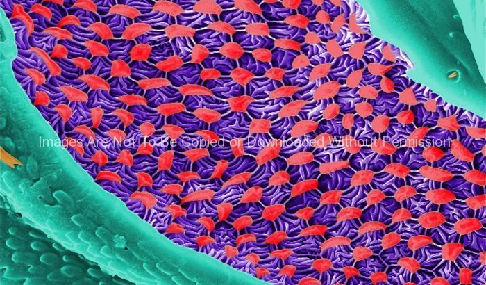
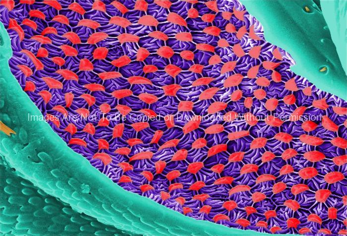
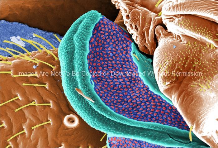
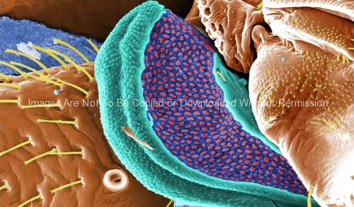
Ultrastructural Morphology Displayed on the Ventral Surface of a Bedbug (Cimex lectularius)
This digitally-colorized scanning electron micrograph (SEM) revealed some of the ultrastructural morphology displayed on the ventral surface of a bedbug, Cimex lectularius. From this view, at the top, you can see the insect’s skin piercing mouthparts it uses to obtain its blood meal, as well as a number of its disarticulated six jointed legs. You’ll also notice a beautiful diaphanous structure at the bottom of the image. It is speculated that this wondrous ultrastructural organ is most probably a scent gland, or related to the dissemination of scent, which may be pheromonal in nature. A further dissection of this, and the adjacent mesothoracic region, could possibly reveal an internalized aspect of this organ, which would be glandular in nature, and actually involved in the production of the aromatic chemical.
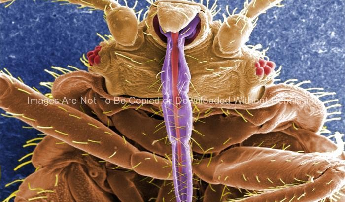
Skin Piercing Mouthparts of a Bedbug, (Cimex lectularius)
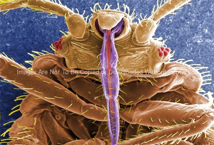
This digially-colorized scanning electron micrograph (SEM) revealed some of the ultrastructural morphology displayed on the ventral surface of a bedbug, Cimex lectularius. From this view you can see the insect’s skin piercing mouthparts it uses to obtain its blood meal, as well as a number of its six jointed legs.
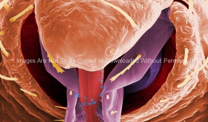
Ultrastructural Morphology on the Rostral Head Region of a Bedbug (Cimex lectularius)
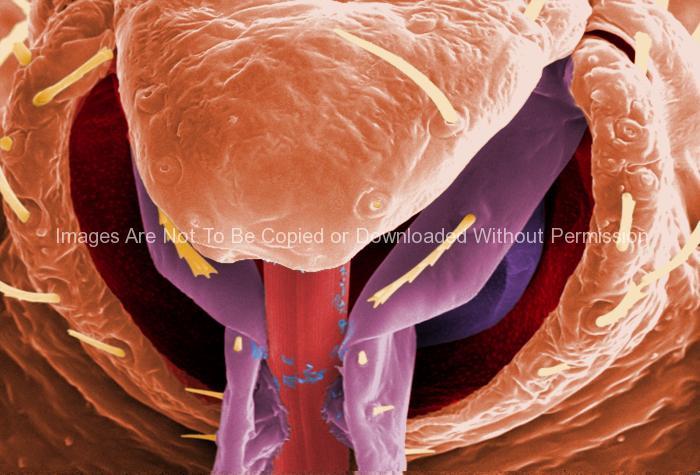
This highly-magnified, digitally-colorized scanning electron micrograph (SEM) revealed some of the ultrastructural morphology displayed on the rostral head region of a bedbug, Cimex lectularius. Note the proximal anatomical relationships the insect’s skin piercing mouthparts it uses to obtain its blood meal, and how they join the head.
