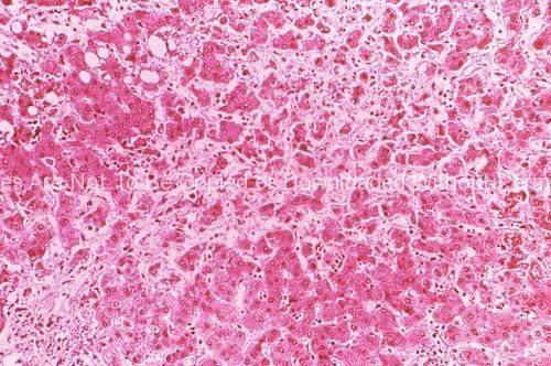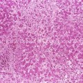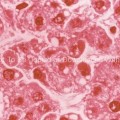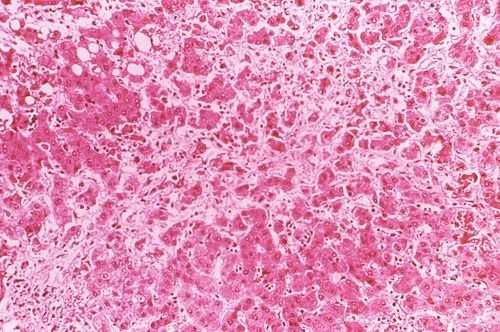
Under a magnification of 100X, this hematoxylin-eosin-stained (H&E) photomicrograph depicts the cytoarchitectural changes found in a liver tissue specimen extracted from an Ebola disease patient in Zaire. This particular view reveals a “zone of acidophilic necrosis within which there were several degenerating hepatocytes. Taking place at the periphery of the necrotic area, were microvacuolar fatty changes.”
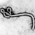
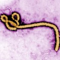
keywords: ebola virus stock photo, virion, micrograph, TEM
