This photograph captures a sneeze in progress, revealing the plume of salivary droplets as they are expelled in a large cone-shaped array from this man’s open mouth, thereby, dramatically illustrating the reason one needs to cover hios/her mouth when coughing, or sneezing, in order to protect others from germ exposure.
Man Sneezing Germs Shown Spreading
This photograph captures a sneeze in progress, revealing the plume of salivary droplets as they are expelled in a large cone-shaped array from this man’s open mouth, thereby, dramatically illustrating the reason one needs to cover hios/her mouth when coughing, or sneezing, in order to protect others from germ exposure.

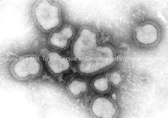
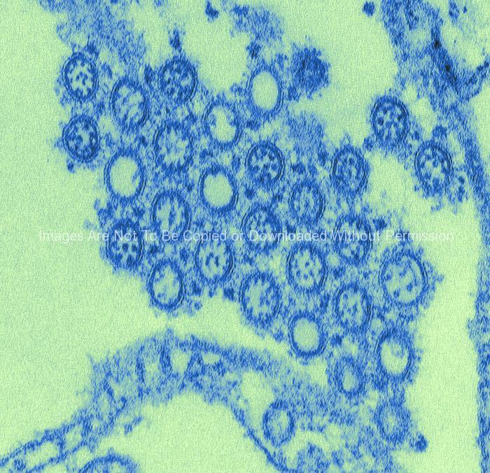
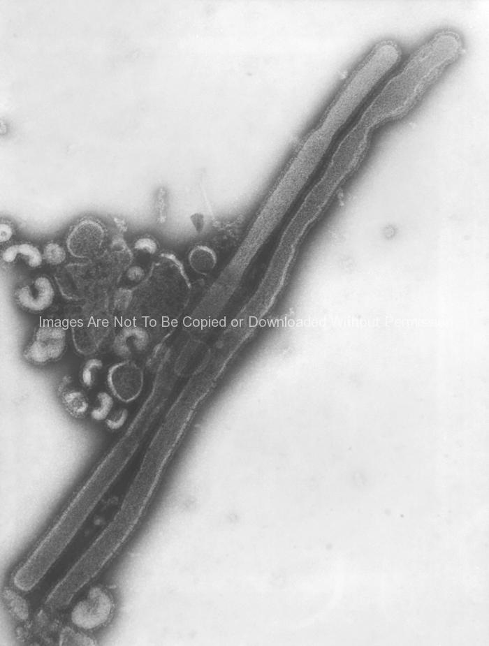
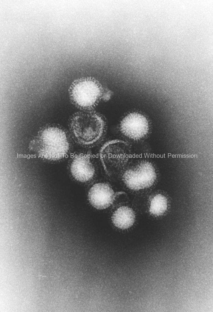
Hong Kong Flu Virus Virions (H3N2 Subtype)
(All Images are for Editorial Use Only)
This negatively-stained transmission electron micrograph (TEM) revealed the presence of a number of Hong Kong flu virus virions, the H3N2 subtype of the influenza A virus. This virus is a Orthomyxoviridae virus family member, and was responsible for the flu pandemic of 1968-1969, which infected an estimated 50,000,000 people in the United States, killing 33,000. Note the proteinaceous coat, or capsid, surroundind each virion, and the hemagglutinin-neuraminidase spikes, which differ in terms of their molecular make-up from strain to strain.
There are many different subtypes of type A influenza viruses. These subtypes differ because of changes in certain proteins on the surface of the influenza A virus (hemagglutinin [HA] and neuraminidase [NA] proteins). There are 16 known HA subtypes and 9 known NA subtypes of influenza A viruses. Many different combinations of HA and NA proteins are possible. Each combination represents a different subtype. All known subtypes of influenza A viruses can be found in birds.