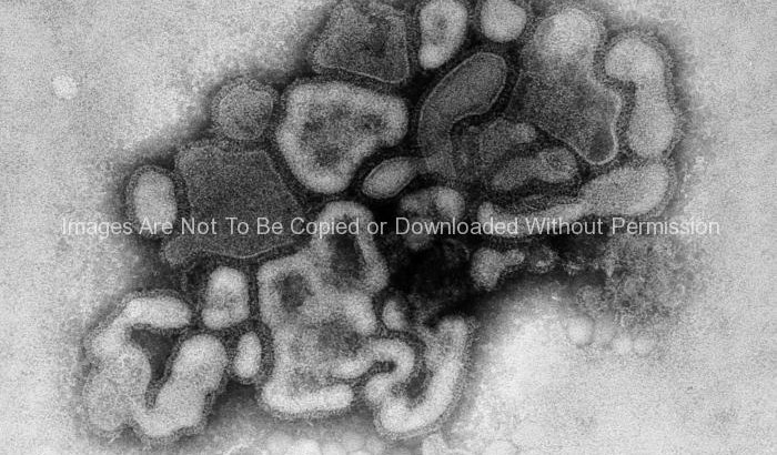
Under a plate magnification of 37,800X, this transmission electron micrograph (TEM) depicted the A/New Jersey/76 (Hsw1N1) virus, while in the virus’ first developmental passage through a chicken egg.
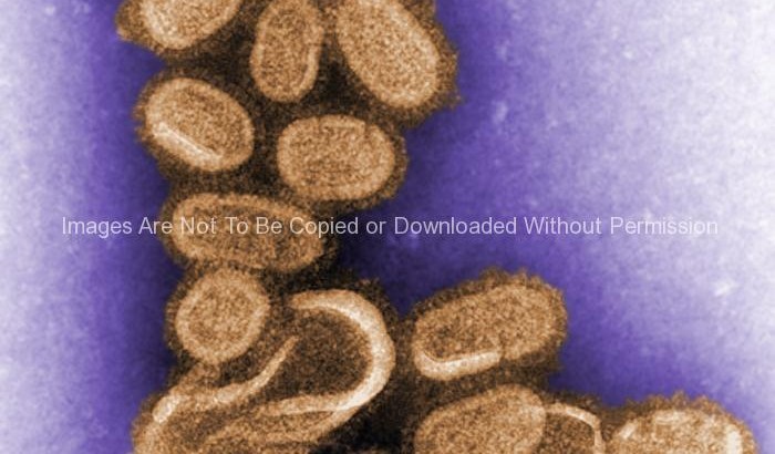
1918 Influenza Virions
This negative stained transmission electron micrograph (TEM) shows recreated 1918 influenza virions that were collected from supernatants of 1918-infected Madin-Darby Canine Kidney (MDCK) cells cultures 18 hours after infection.
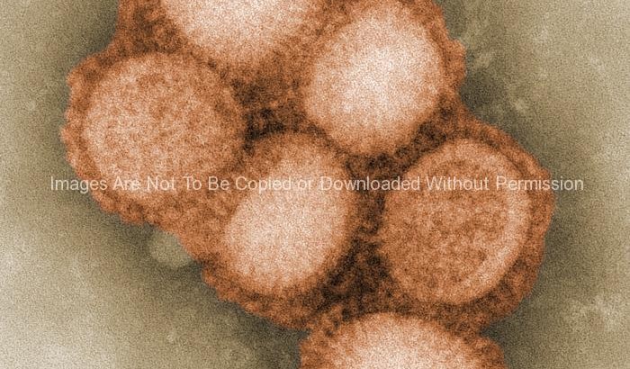
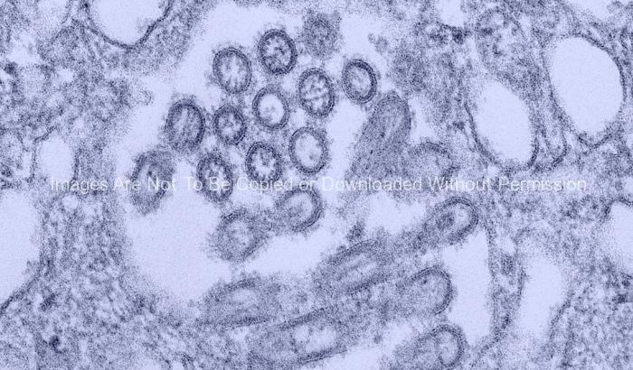
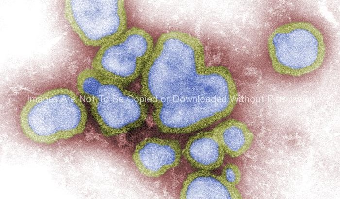
Morphology of the A/CA/4/09 Swine Flu Virus
This colorized negative stained transmission electron micrograph (TEM) depicted some of the ultrastructural morphology of the A/CA/4/09 swine flu virus.
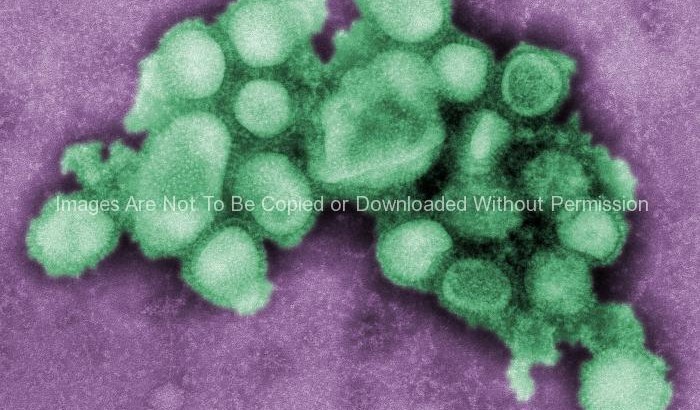
Morphology of the A/CA/4/09 Swine Flu Virus
This colorized negative stained transmission electron micrograph (TEM) depicted some of the ultrastructural morphology of the A/CA/4/09 swine flu virus.
