This transmission electron micrograph (TEM), taken at a magnification of 150,000x, revealed the ultrastructural details of an avian influenza A (H5N1) virion, a type of bird flu virus which is a subtype of avian influenza A. At this magnification, one may note the stippled appearance of the roughened surface of the proteinaceous coat encasing the virion.

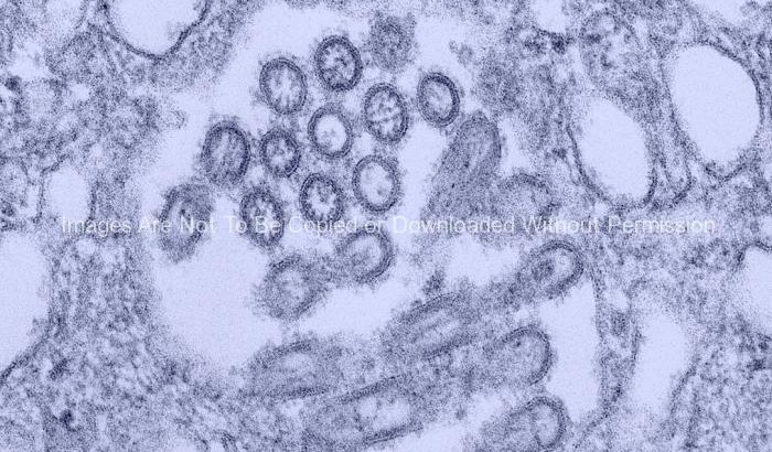
Russian Influenza-A H1N1, (A/USSR/90/77 strain)
This transmission electron micrograph (TEM) depicted Russian influenza-A H1N1, (A/USSR/90/77 strain), virions, which had been magnified 189,000x.
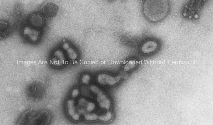
Russian Influenza-A H1N1, (A/USSR/90/77 strain)
This transmission electron micrograph (TEM) depicted Russian influenza-A H1N1, (A/USSR/90/77 strain), virions, which had been magnified 189,000x.
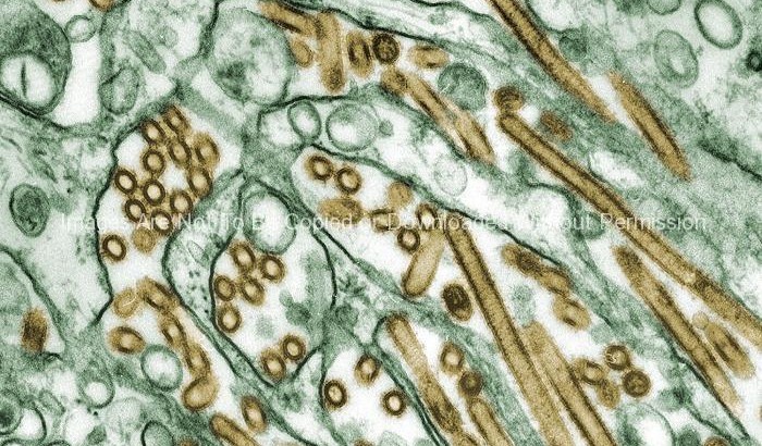
Electron Micrograph of Avian Influenza A
Colorized transmission electron micrograph of Avian influenza A H5N1 viruses (seen in gold) grown in MDCK cells (seen in green).
Avian influenza A viruses do not usually infect humans; however, several instances of human infections and outbreaks have been reported since 1997. When such infections occur, public health authorities monitor these situations closely.